What Does An Animal Cell Look Like

Let’s be honest—thinking about what an animal cell looks like might sound like a school biology flashback. But it’s actually one of those fascinating subjects that connects to...
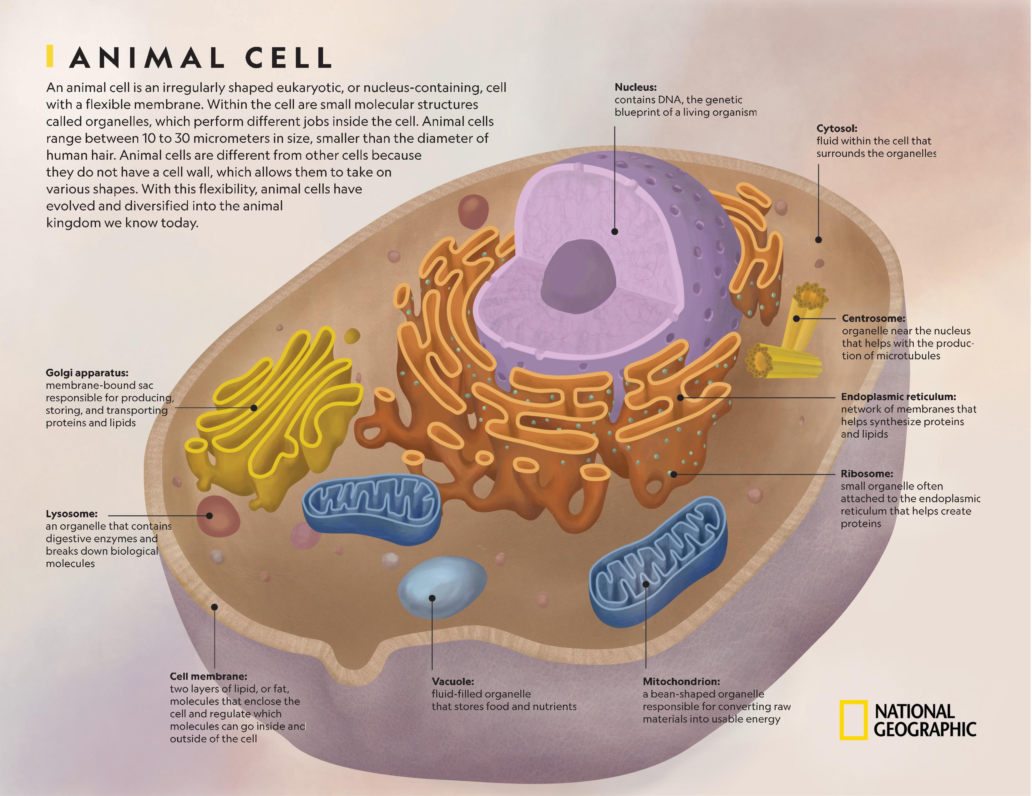
Let’s be honest—thinking about what an animal cell looks like might sound like a school biology flashback. But it’s actually one of those fascinating subjects that connects to everyday life. Ever wonder how your muscles heal after a workout, or why a bee sting hurts so much? That’s your cells talking. Understanding the basic shape and parts of an animal cell is like peeking under the hood of a car you drive every day: your own body. It’s practical for students cramming for a test, curious hobbyists, or anyone who wants to appreciate the incredible machinery that makes us tick.
An animal cell isn’t a perfect circle or square—it’s more like a soft, squishy bag that can change shape. Unlike plant cells, which have a rigid wall, animal cells have a flexible outer layer called the cell membrane. Think of it as a security gate that lets good stuff in (like sugar) and kicks bad stuff out (like waste). Inside, it’s a busy jelly-like substance called cytoplasm, where all the action happens. If you imagine a tiny, bumpy water balloon filled with floating gems, you’re on the right track.
Now, let’s highlight the star players. Floating in that cytoplasm is the nucleus—a dark, round blob that’s the cell’s brain. It holds your DNA, the instruction manual for everything your body does. Then there are mitochondria, often called the power plants. They look like little bean-shaped capsules and are responsible for turning food into energy. If you’ve ever felt a burst of energy after a good meal, thank these tiny guys. They’re so crucial that muscle cells are packed with them.
Must Read
You’ll also spot smaller structures called ribosomes (tiny dots) and the endoplasmic reticulum (a wrinkly, folded membrane). Imagine a factory conveyor belt—that’s the ER, making and shipping proteins. Ribosomes are the workers on that belt. And there’s the Golgi apparatus, which looks like a stack of pancakes. It packages proteins and sends them out to other parts of the body. For example, when you get a cut, these structures help deliver healing materials to the wound. Variations exist too: a red blood cell looks like a donut to squeeze through tiny vessels, while a nerve cell is long and spiky, like a tree root.
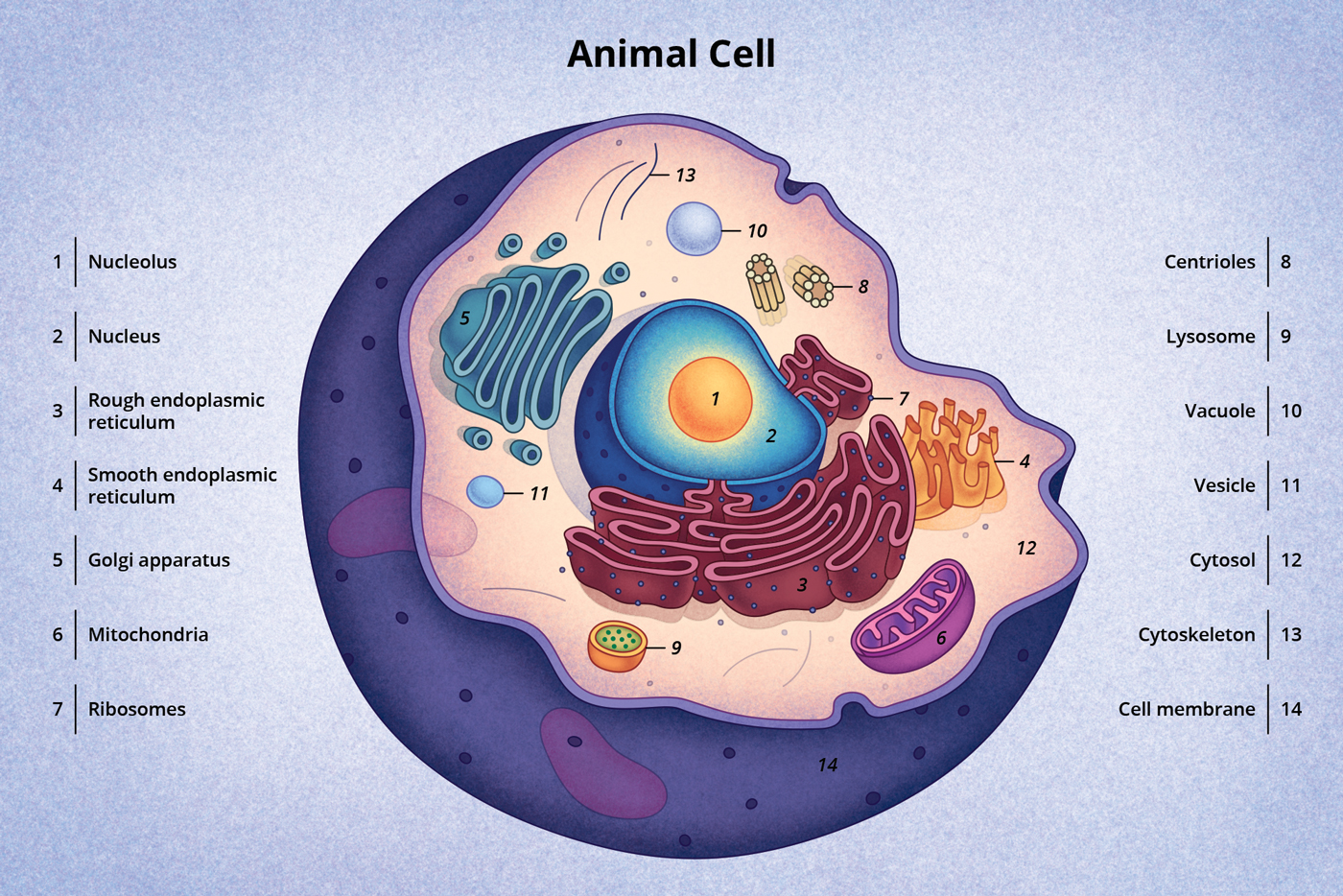 Animal Cell Labeled Diagram – Animal Cells Diagram – IMCUI
Here’s a simple tip to picture it: take a handful of Jell-O with fruit pieces in it. The Jell-O is the cytoplasm, a chunk of pineapple is the nucleus, blueberries are mitochondria, and a few sprinkles of granola represent ribosomes. This mental model makes it easy to remember. Another trick: draw a circle, then add a few blobs inside—don’t stress about perfect shapes. Nature is wobbly and imperfect, and that’s exactly how a real cell looks.
To get the most out of this, start with one structure at a time. Look at a picture of an animal cell online, then cover it and try to sketch from memory. Focus first on the nucleus and mitochondria—those are the easiest to spot. For a deeper appreciation, watch a time-lapse video of a cell dividing. You’ll see the membrane wobble and the nucleus break apart, proving that these “bags” are alive and constantly moving. That’s the real beauty: an animal cell is not a diagram in a textbook, but a dynamic, squiggly little world that keeps you breathing, moving, and thinking.
/animal_cell-56c765663df78cfb3788382b-5c2e861046e0fb000142aa47.jpg)

